🩺 Case Overview
Patient Information
- Breed: Poodle
- Sex: Male
- Age: 6 years old
- Weight: 4 kg
The patient, a 6-year-old male Poodle, suffered a right forelimb injury when a delivery tricycle ran over him during a walk. The dog showed severe pain, swelling, and inability to bear weight. Radiographs confirmed distal radial and ulnar fractures.
This was a high-energy trauma fracture in a small breed, requiring a fixation method that maintained stability while minimizing soft tissue and blood supply damage.
🦴 Surgical Treatment
X-rays showed:
- Comminuted distal fracture of the radius
- Transverse distal fracture of the ulna
Given the bone structure and vascular fragility of small-breed dogs, the surgical plan included:
Radial plate fixation with conservative healing of the ulna.
This approach preserved periosteal integrity, minimized tissue trauma, and provided stable alignment for effective bone healing.

⚙️ Surgical Procedure
Under general anesthesia, the surgical area was aseptically prepared. An incision was made along the medial side of the radius to expose the fracture. The periosteum was carefully separated while protecting nearby vessels and nerves.
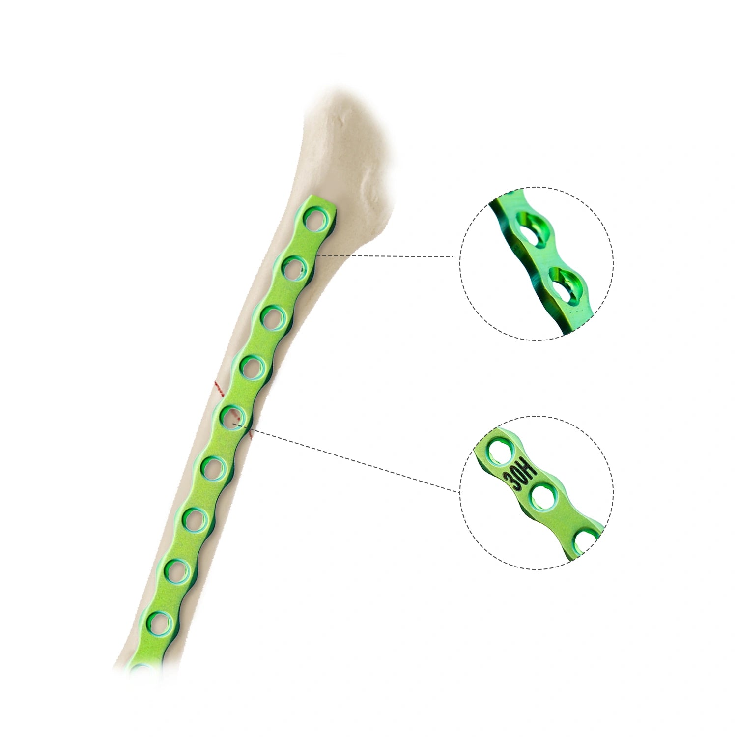
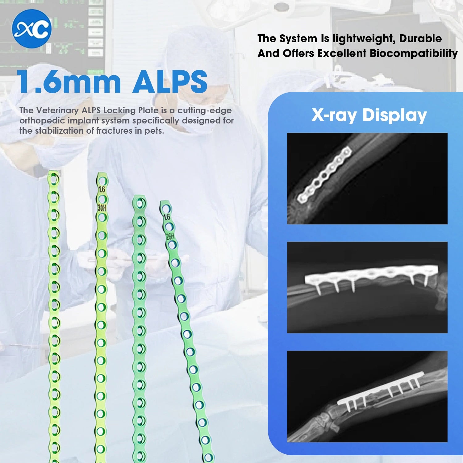
After reduction and alignment of the fracture ends, a small LCP plate was positioned on the palmar aspect of the distal radius. Screw holes were drilled at a safe distance from the fracture line, using continuous saline irrigation to remove bone debris.
Once fixation was achieved, stability and joint mobility were checked — ensuring smooth movement without friction.
An external splint was applied postoperatively to assist internal fixation, followed by radiographic confirmation of proper alignment and implant stability.
💊 Postoperative Care
Medication:
The patient received intravenous therapy of 0.9% saline (50 mL), ceftriaxone sodium (20 mg/kg), and butorphanol (0.1 mL) for 7 days.
Daily wound disinfection with enzymatic spray maintained a sterile environment.
Home Care:
An Elizabeth collar prevented licking and self-trauma. The dog was kept in a soft, clean crate to limit movement.
Diet included digestible food, calcium, and cod liver oil to support bone recovery, with adequate hydration.
💪 Recovery & Follow-Up
The first week focused on rest and wound healing. Sutures were removed after 7 days, followed by gentle passive stretching exercises to improve limb mobility.
By week two, controlled walking was introduced to prevent muscle atrophy.
At one month, X-rays showed proper healing with stable fixation. At two months, bone callus formation was evident with no misalignment.
By three months, the dog had regained normal walking ability without lameness.
Rehabilitation Tips:
- Avoid jumping and running for at least three months.
- Encourage short, frequent walks to rebuild muscle strength.
- Schedule X-ray rechecks to ensure complete bone union.
📋 Conclusion
This case highlights the effectiveness of combined internal and external fixation for treating small-breed forelimb fractures.
Protecting soft tissue and maintaining blood supply were key to successful bone healing.
The combination of precise surgical technique, stable fixation, and attentive postoperative care led to excellent recovery with full limb function.
- WhatsApp:+86 173 1508 9119


